Stroboscopic Epifluorescence Intravital Microscope This is a Zeiss Axioscope upright microscope. It has a DC brightfield (suitable for velocity measurements) and fluorescence illuminator. Fluorescence illumination is powered by a xenon strobe that is triggered by the video signal (60 flashes per second). Filters suitable for GFP, RFP, fluorescein and rhodamine are available. 20X 0.50NA, 40X 0.75NA and 63X 0.90NA water immersion objectives are mounted. Recording is analog through a high-sensitivity DAGE SIT66 camera into a VCR. For analysis, video is piped into a frame grabber. Each frame has a time stamp. A second, very similar microscope is equipped with a high-resolution digital camera (Sensicam QE), which provides much better spatial resolution at the expense of a slightly lower frame rate. Applications Intravital experiments This upright microscope has water immersion objectives with long working distance, large approach angles for manipulator access, which is ideal for intravital experiments. The extremely short illumination achieved by the strobe (microseconds) beats any shuttered camera. This microscope is optimized for imaging fast-moving objects like leukocytes and platelets in free-flow or rolling. Since light toxicity is essentially absent, transmigration studies can also be done. Dual imaging The camera is monochrome (black and white), so only one color can be recorded at a time. However, mixed chimeras where some cells express GFP and others not are very suitable (figure 1) for side-by-side comparison of knockout (typically appear grey) and wild-type cells (appear white when GFP is used) in the same microvessel. Also suitable for transillumination microscopy (figure 2) and micro-flow chamber work (figure 3). Location: Animal facility room 8. Contact information: Javier Mestas, IB-Ley lab Training procedures contact information: Leo Fernandez 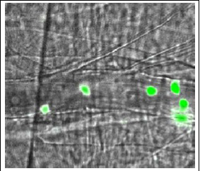 Figure 1. Neutrophils from two different donor mice rolling in the same cremaster muscle microvessel. A lethally irradiated wild-type mouse was transplanted with a mixture of bone marrow cells from lysozymeM-GFP mice (resulting in green neutrophils) and from Tyrobp-/-Fcrg-/- double knockout mice (resulting in unlabeled neutrophils, grey). Note that white was converted to green by image processing. From Zarbock and Ley, JEM 2008 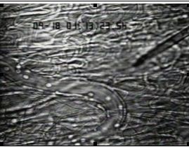 Figure 2. Rabbit neutrophils rolling and arresting in response to IL-8 (injected into tissue by micropipette, from right). From Ley et al., JI 1993. 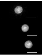 Figure 3. Mouse neutrophil rolling (three successive frames) in a whole-blood perfused flow chamber on E-selectin. The neutrophil expresses GFP under the lysozyme M promoter. Stroboscopic epifluorescence with Sensicam QE. Setup similar to Chesnutt et al., Microcirculation 2006. |
|||||||||||||||||||||||||||||||||||||||||||||||||||